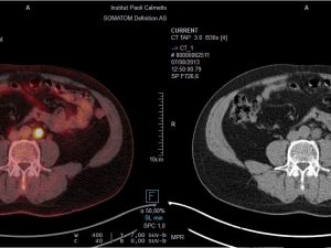
The role of imaging techniques such as multiparametric Magnetic Resonance Imaging (MP-MRI) will expand in the next few years and will have a more central role in the diagnosis of prostate cancer (PCa), particularly in cases when new technologies are combined to yield more detailed images.
“MP-MRI will play an important role in guiding both biopsy as well as focal therapy,” said radiologist Dr. Jurgen Futterer of the Radboud UMC in Nijmegen (NL). Futterer will lecture during the upcoming 4th Meeting of the EAU Section of Urological Imaging (ESUI) on November 12 in Barcelona, Spain. The 4th ESUI will be held in conjunction with the 7th European Multidisciplinary Meeting on Urological Cancers (EMUC).
“MP-MRI can detect clinical significant cancers in males with rising PSA and negative biopsies. The negative predictive value is around 95% (this is based on a recent systematic review in European Urology). However, non-significant cancers may be missed (Gleason 6 cancers). MP-MRI can be applied in patients who are opting for active surveillance and active treatment follow-up,” explained Futterer when asked how doctors can achieve optimal treatment results.
“Moreover, MP-MRI can be used to select patients for focal therapy,” he added while underscoring that optimal PCa treatment will increasingly rely on imaging results and capabilities.
Futterer will lecture on the topic “What is the new standard of prostate MRI? Mandatory sequences and PIRADS 2.0.” The lecture is part of a Point-Counterpoint session where experts will debate on whether MRI detects significant prostate cancers. ESUI Chairman Jochen Walz (FR) will present counter arguments while Dr. H.U. Ahmed (GB) would argue in favor of MRI.
Futterer will discuss the essential imaging sequences (T2-weighted anatomical imaging, diffusion weighted imaging and contrast enhanced MR imaging) in his lecture, highlighting the role of each procedure. He will also look into the role and application of PI-RADS v2, the second and revised version of Prostate Imaging-Reporting and Data System (PI-RADS) which refers to a structured reporting scheme for prostate cancer.
With the emphasis on sophisticated imaging techniques in recent years, Futterer said he expects to see new developments which will impact PCa diagnosis.
“Most likely diffusion weighted imaging will evolve into a biomarker for cancer aggressiveness. Furthermore, finger printing imaging techniques will be available in which from one MR imaging sequence acquisition we will able to extract different tissue contrast,” he said.
Before this happens, however, more research into the exact role of imaging needs to be done, particularly in the multi-disciplinary setting where various prostate cancer experts are involved in the treatment management.
In current guidelines, according to Futterer, MP-MRI is recommended in certain indications. “I think that imaging has a more prominent role,” he noted given the fact the doctors need a tool that will allow them to draw more informed conclusions, enabling optimal treatment strategies.
Regarding challenges in the field of imaging for urological cancers, he noted the role of prostate-specific membrane antigen (PSMA) PET imaging in detecting lymph node metastasis. “Currently imaging is limited for lymph node metastases,” he said implying that experts need to further look into the role of PSMA.
The ESUI Meeting has prepared an exciting programme which will cover the most recent developments in urological imaging, with topics up for discussion and debate in a number of roundtable sessions. The day-long agenda will tackle individualised medicine, new imaging technologies on the horizon, molecular imaging (in a joint session with the European Association of Nuclear Medicine) and optimising PC a management, among other topics.


