In a rainy and rather cold day in November last year, the 5th meeting of the EAU Section of Urological Imaging (ESUI16) took place in Milan, Italy, a day before the 8th European Multidisciplinary Meeting on Urological Cancers (EMUC).
ESUI16 marked the third time the ESUI meeting has been organised in conjunction with EMUC and other activities such as the European School of Urology (ESU) courses, the EMUC Symposium on Genitourinary Pathology and Molecular Diagnostics (ESUP) and the EAU Young Academic Urologists (YAU) Meeting.
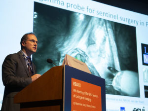
The meeting’s success was remarkable due to the high audience number and a high level of interaction among the speakers and participants (Figure 1). The main topics has been divided in three main sessions, namely: 1) Imaging with multiparametric MRI (mpMRI) and target biopsies 2) Molecular imaging in urology (Joint session of ESUI and EANM), and 3) Improving imaging for urothelial cancer.
Role of mpMRI and target biopsy
mpMRI combining T2-weighted, diffusion-weighted, and contrast-enhanced dynamic imaging is recognized as the most accurate imaging modality for prostate cancer detection and characterization. As everybody has anticipated, mpMRI of the prostate has become an exploding phenomenon that can no longer be ignored. Even if mpMRI has a strong indication only before a second biopsy (see EAU Guidelines), speakers N. Mottet (FR) and H.U. Ahmed (GB) have both supported the use of mpMRI before the primary biopsy in their point-and-counterpoint discussion in prostate cancer diagnosis. The conclusion was that, despite the costs which are still high, mpMRI will be used more in the future.
In the same session, the presentation of T. Cai (IT) on how to overcome the problem of sepsis prompted a long and interesting discussion, even if the problem of the higher rate of sepsis remains unresolved.
Subsequently, A. Postema (NL), G. Salomon (DE) and M. Ritter (DE) presented interesting insights on multiparametric ultrasound, new ultrasound-based technologies and the various weapons or tools when performing a biopsy, respectively.
In the replay session (called ‘How I Do It’) of pre- recorded videos, various speakers have presented different technologies. The conclusion of the session was that, like any valuable new technology, targeted prostate biopsy is evolving rapidly, and current methods would certainly improve in the near future.
Refinements in MRI are in the horizon, and radiologists trained to perform and interpret prostate MRI will increase in number. Industry-driven improvements in image-fusion hardware and software are also expected due to the large market potential for improving prostate biopsy. Image fusion devices will also become smaller and easier to use. Co-registration accuracy will increase, and graphical user interfaces will become more intuitive. Already, targeted prostate biopsy has a serious online visibility, and various support groups are making it a priority item.

Photo: Joana Neves receives the ESUI Best Poster Award
Attempts to image CaP via modalities simpler than MRI (e.g., elastography, contrast-enhanced ultrasound (US), high frequency US, Anna/c-TRUS) have been shown during the meeting by other renowned experts. Following the session Joana Neves (UK) won the ESUI Best Poster Award with her study titled “Combining mpMRI sequences for the diagnosis of prostate cancer – the value of adding diffusion and contrast enhancement to T2W on 3Tesla: Outcomes from the PICTURE Trial” (See Box, Figure 2).
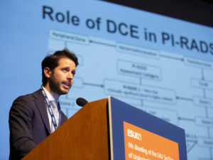
During the afternoon session on changing paradigms in prostate cancer treatment, J. Walz (FR) and the author tackled the hot topic of mpMRI and active surveillance (AS) in cases of a visible lesion. Both speakers concluded that mpMRI and target biopsies have important roles in selecting and following men on AS. Progression on mpMRI may predict the risk of pathological progression and patients with stable imaging have a low rate of progression. Ideally, as imaging and target biopsy technology evolve the number or frequency of biopsies in men on AS may potentially be reduced. Furthermore, mpMRI may allow for expanded AS criteria, permitting more men to participate in AS with higher confidence in accurate monitoring of disease progression (Figures 3-4).
G. Thalmann (CH) discussed the pilot research into focal therapy, using technologies derived from image-guided biopsy. Meanwhile, A. Villers (FR) and J. Futterer (NL) discussed the training of urologists and radiologists on how to read a prostate mpMRI and the expertise needed in this area, respectively.
Molecular Imaging in Urology: Joint ESUI-EANM Session
As done in previous years, ESUI16 held a joint ESUI and European Association of Nuclear Medicine (EANM) session that focused on two main themes: the clinical utility and pitfalls of nuclear medicine imaging in urological oncology, and the use of Prostate specific membrane antigen- positron emission tomography (PSMA PET) in prostate cancer.
All speakers were unanimous in saying that, to date, the biggest contribution of nuclear medicine to urological oncology has been to aid in lymph node and distant disease staging and that, for prostate cancer, PSMA PET appears to perform better than other nuclear medicine imaging modalities. Despite this, while PSMA PET is able to detect prostate cancer nodal recurrence earlier than other imaging modalities, doubts still exist as to whether any change in clinical management at this point is warranted or beneficial.
Alberto Briganti (IT) revealed recent analyses by his group which showed that, when compared to extended bilateral salvage pelvic and retroperitoneal lymph node dissection, PSMA PET scan can underestimate the burden of nodal metastasis and may not be appropriate to guide limited dissection. Lars Budaus (DE) also presented evidence that micrometastatic disease (below the 5mm threshold) can be overlooked by PSMA PET in prostate cancer.
Tobias Maurer (DE) presented potential uses of PSMA PET, such as targeted radiation therapy, and also disclosed preliminary results on the use of a PSMA intracorporeal gama probe to guide lymph node dissection.
Stefano Fanti (IT) (stepping in for F. Giesel [DE]), presented Giesel’s early data suggesting a good diagnostic performance of PSMA PET in early, localised prostate cancer. The use of fluorinated PSMA (18F-PSMA-1007) instead of gallium could be a game changer as this tracer has no urinary excretion, allowing for better assessment of the bladder and eliminating the production of radioactive urine, which can contribute to a more widespread use. In his own lecture, Fanti also mentioned other new tracers in prostate cancer such as the anti-1-amino-3-18F- fluorocyclobutane-1-carboxylic acid or FACBC and the recently FDA-approved 18F-fluciclovine, and the possible uses of molecular imaging to assess clinical response to systemic therapies such as docetaxel.
Antoni Vilaseca Cabo (ES), the only speaker to move away from prostate, discussed the clinical usefulness of molecular imaging in bladder cancer. Computerised tomography (CT) is still the gold standard for nodal and distant disease staging in bladder cancer and the use of choline or FDG PET CT do not seem to improve on the diagnostic ability of CT alone. However, in the near future, evidence may show that PET MRI supplants CT. In the more distant future, molecular imaging radiomics may be the key to improve staging in bladder cancer.
In conclusion, molecular imaging has contributed and is envisioned to continue to contribute to advances in urological oncology. PSMA PET’s most consensual use is still as an end-of- line imaging tool in prostate cancer, when clinical queries persist and more definitive answers are needed to change clinical management. New tracers and the combination of PET and MRI can impact the diagnostic setting but efforts to produce more robust and consistent evidence on the benefits of molecular imaging should be sought out.
Improving imaging for urothelial cancer
This session mainly focused on technologies to improve urothelial cancer in the bladder and in the ureter by photodynamic diagnosis (PDD) versus Narrow Band Imaging (NBI) versus Storz professional image enhancer system (SPIES) or Optical coherence tomography (OCT), as nicely shown by B. Geavlete (RO) and M. Bus (NL). Even if they are not really new instruments, these technologies had reached a very high level of visualisation and they are considered all good alternatives. Last but not least, N. Cowan (GB) has shown the importance of the perfect Uro-CT in the detection of urothelial cancer and the need for standardisation.
The 5th ESUI Meeting has proven to be a successful meeting with excellent speakers and a responsive audience who actively participated in the discussions. The Scientific Programme covered all the major topics in urological imaging and this achievement is a sound basis for the next meeting in Barcelona, Spain in November this year.
Authors
Dr. Vincenzo Scattoni, Dept. of Urology Ospedale San Raffaele
University Vita-Salute Milan (IT)
Dr. Joana Neves, Division of Surgery and Interventional Science
University College London, London (GB)